Bones
Bones have many shapes and sizes and are important to add structure to the body and protection to the vital structures. The bones have a crystalline construction embedded with mineral and live cells that maintain and repair the skeleton.
Jump to:
Shoulder Bones
 Scapula
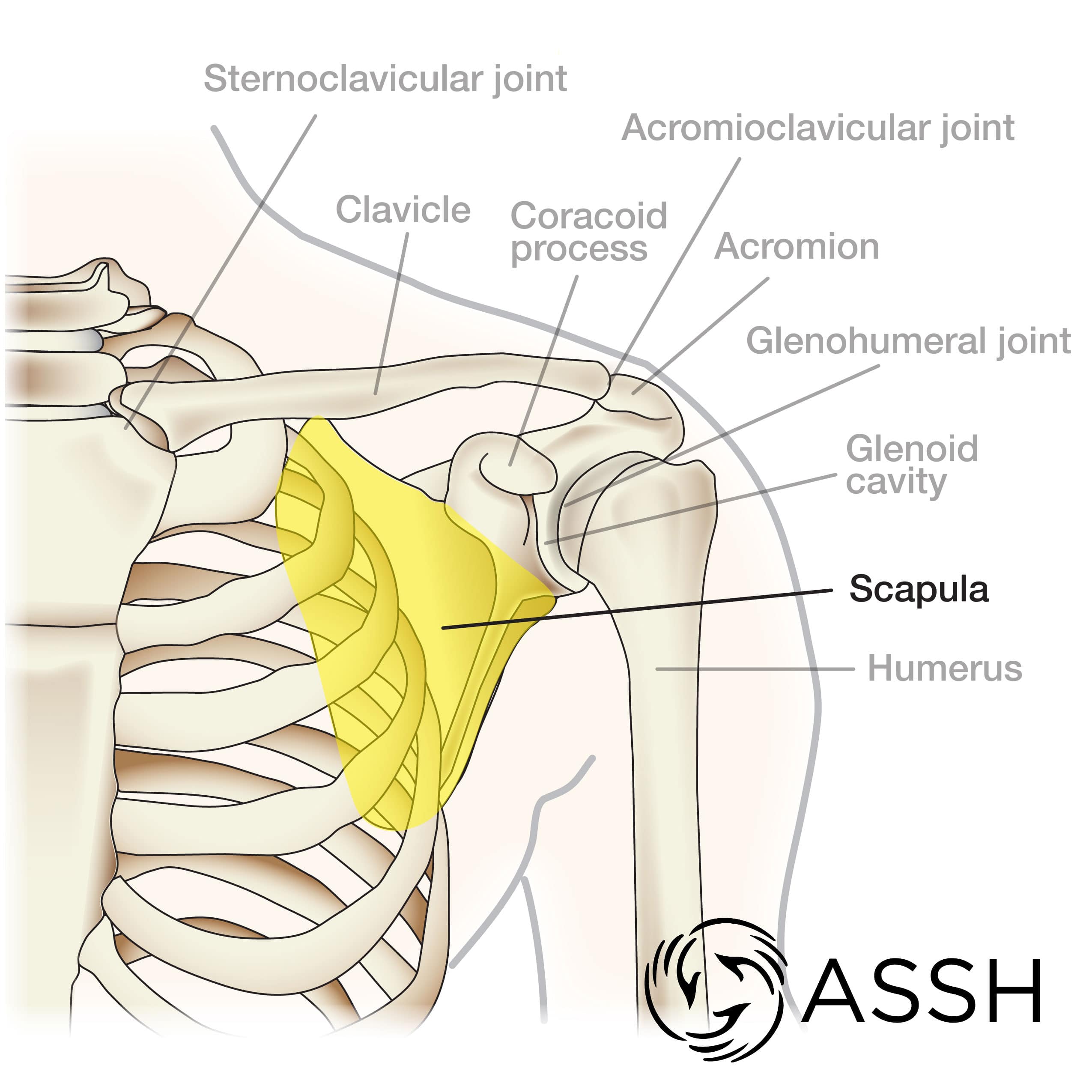
The scapula, or “shoulder blade,” is an approximately triangular shaped bone. It, essentially, floats off of the back of the chest, as it is connected to the body primarily by muscle. In fact, there are 17 muscles that attach to the scapula. The scapula has a joint that wraps around from the back to the front of the shoulder called the acromion. The acromion is the part of the scapula that attaches to the collar bone and is the only true joint attaching the arm to the body. Much of the motion of the shoulder is actually motion between the scapula and the chest. Because of this, the main shoulder joint (called the glenohumeral joint), the scapula, and the surrounding muscle and ligament are collectively referred to as the shoulder girdle. The shoulder girdle combines to give you shoulder motion. Injuries to the scapula are usually from an awkward fall or car accident.
Clavicle
The clavicle, or “collar bone,” is a long slightly curved bone that connects the arm to the chest. In most people, the clavicle is easy to feel and even see under the skin. The clavicle attaches to several muscles connecting it to the arm, the chest and the neck. There are two ends with joints on the clavicle, and these can become arthritic in some people. Clavicle fractures typically happen after a fall or other significant trauma.
Acromion
The acromion is a fairly flat projection of the scapula that curves around from the back to the front of the shoulder. Much of the strong deltoid muscle around the shoulder attaches to the acromion. This bone also gives the shoulder much of its almost squared-off shape. In some people there is an extra piece of bone that, during development, did not fuse to the rest of the acromion. This is called an os acromiale. Occasionally the os acromiale can cause discomfort. However, most frequently it does not cause a a problem. Lying under the acromion, there is a layer of bursa tissue and some of the rotator cuff. Some people have a curve or hook to the underside of the acromion near the rotator cuff tendons. Surgeons sometimes suggest removal of the curve or hook at the time of surgery.
Coracoid Process
The coracoid process is a projection off of the scapula that points straight out to the front of the body. This piece of the scapula bone is important because it has muscles and ligaments attached that help hold the clavicle, the shoulder joint, and humerus. There are ligaments from the coracoid that help keep the clavicle in place; these can be torn in some AC (acromioclavicular) joint dislocations. One of the heads of the biceps attaches to the coracoid. The coracoid usually does not cause pain or have injuries but can occasionally be a cause for shoulder discomfort.
Glenoid cavity
The glenoid is the socket portion of the ball and socket joint of the shoulder (glenohumeral joint) and is part of the scapula. It is relatively flat which allows the joint to be the most mobile joint in the body. The glenoid cavity includes the glenoid’s bone and cartilage surface, and it combines with soft tissues like the glenoid labrum, multiple shoulder ligaments and the joint capsule (the lining of the joint) to make the joint stable. While the joint is normally stable, lots of motion, injury or abnormality in any of the structures of the glenoid cavity can lead to joint instability. Occasionally, the shoulder can lose motion from conditions such as arthritis or adhesive capsulitis (frozen shoulder).
Arm Bones
 Humerus
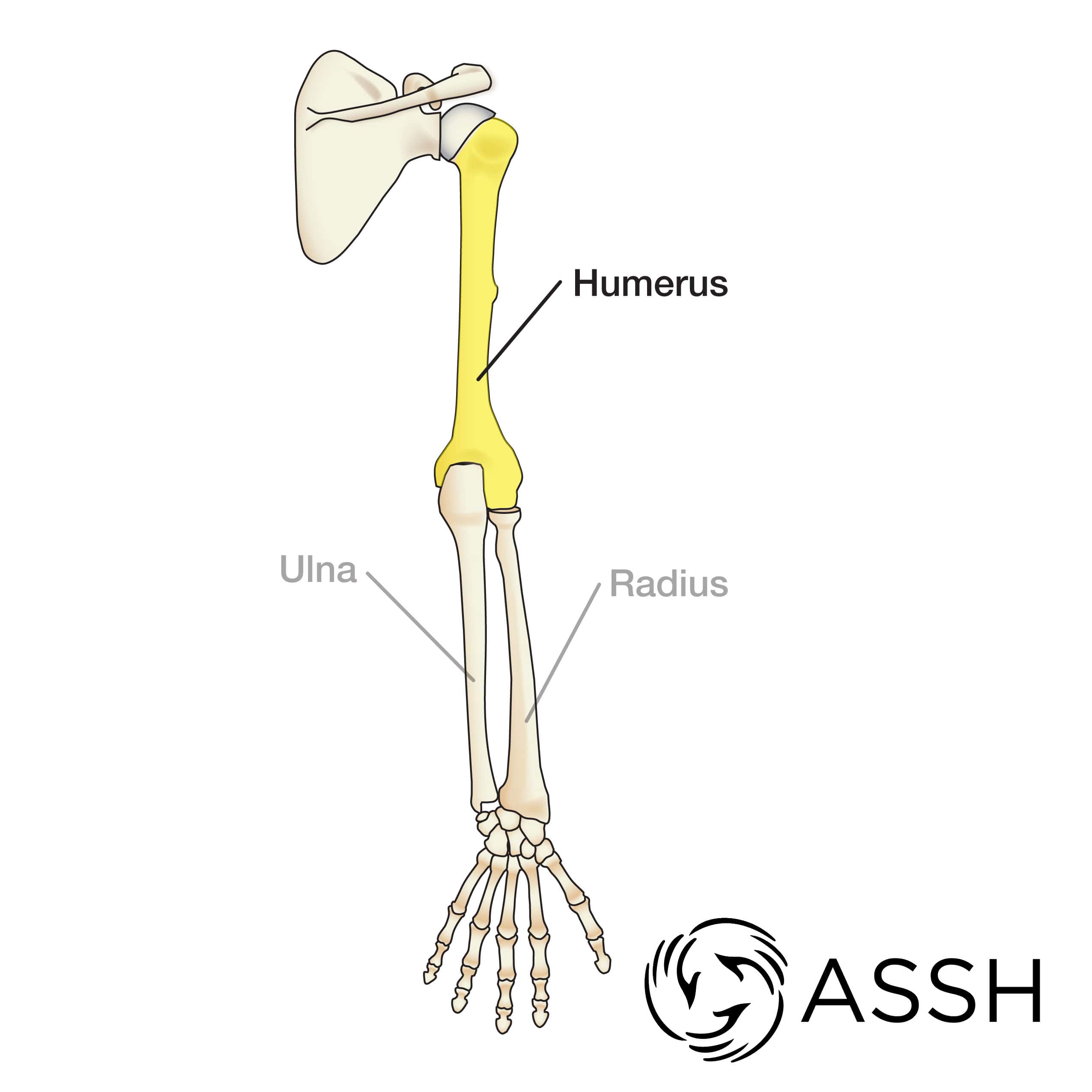
The humerus is the long bone between the shoulder and the elbow. It has the ball of the ball and socket of the shoulder (glenohumeral) joint. At the other end, it has its portion of the elbow joint. The humerus serves as an attachment of many muscles and ligaments in the arm. Some of the attached muscles run all the way into the hand. The humerus typically becomes a problem only when it breaks (fractures). There are many types of humerus fractures and as a result, the treatments for these fractures are quite variable.
Radius
The radius is one of the two forearm bones and is on the thumb side of the forearm near the hand, but is always on the outside of the elbow. The position of the radius changes depending on how the hand is turned because the radius twists around the other forearm bone, the ulna. At the elbow, the radius is part of the odd shaped joint between the humerus and the two forearm bones. The joint between the radius and humerus by itself is like a ball and socket joint, with the radius forming the socket. The radius has many muscular attachments to move the elbow, forearm, wrist and fingers. The end of the radius leads to the wrist joint. The radius and ulna are joined by cartilage joints at the elbow and at the wrist. They are also joined by multiple ligaments. There are many ways that people injure the radius and the forearm. Breaking this bone is common because when we fall, the hands and arms are typically used to break the fall.
Ulna
The ulna is one of the two forearm bones and is on the small finger side of the forearm. Unlike the radius, this bone does not twist, so when the hand changes position, the ulna is always in the same position on the inside part of the forearm. Like the radius, the ulna has joints at the elbow and wrist. The joint between the ulna and humerus is a hinge type of joint. At the wrist, the ulna has a smaller surface in contact with the wrist bones and typically bears less of the force from the hand and wrist. The ulna is joined to the radius throughout the forearm with cartilage joints at the elbow and wrist and multiple ligaments connecting to the radius through the whole length of the forearm. Like the radius, breaking the ulna bone is a common reason for problems with the ulna.
Wrist Bones
 Scaphoid
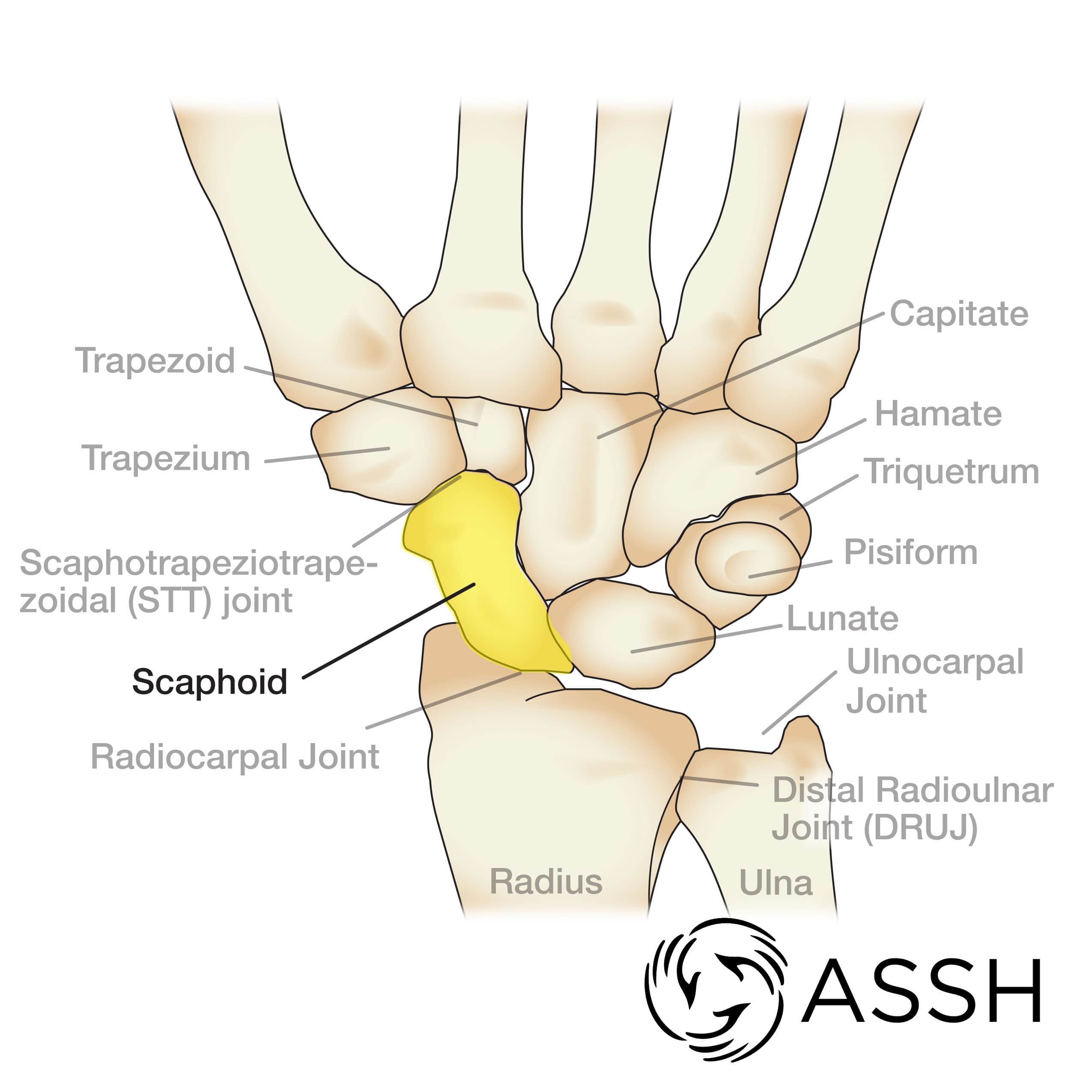
The scaphoid is a bone in the wrist. It is part of the first row of wrist bones, but it helps to link the two rows of wrist bones together. Its name derives from the Greek word for boat because it’s thought that the scaphoid resembles a boat. Most of the scaphoid is covered with cartilage which contacts five other bones in the wrist and forearm. The part of the scaphoid without cartilage is attached to ligaments and has blood vessels that come from the radial artery. Bones need blood flow to heal. A broken or fractured scaphoid can have difficulty healing, or may never heal, because of a disruption of blood flow through the scaphoid. An intact scaphoid is important and necessary for proper wrist function because of how it interacts with the other wrist bones.
Lunate
The lunate is a bone in the middle of the wrist in the first row of wrist bones. Like most of the wrist bones, it is almost entirely covered in cartilage. This bone has a crescent shape when seen from the side and its large cartilage surface allows for significant wrist motion. It is uncommon to break the lunate, but the lunate can be involved with dislocations of the wrist and can rub against the ulna if the ulna is too long compared to the radius bone.
Triquetrum
The triquetrum is a bone on the small finger side of the wrist in the first row of wrist bones. This bone adds stability to the wrist, gives the wrist a larger surface to bear weight transmitted from the hand, and makes a joint with other carpal bones including the pisiform.
Trapezoid
This is a roughly trapezoidal-shaped bone in the second row of wrist bones and primarily holds the index finger metacarpal bone in place. This bone is uncommonly injured.
Trapezium
The trapezium is a saddle-shaped bone in the second row of wrist bones, and it is the main place where the thumb metacarpal connects to the wrist. This bone has an odd shape that allows the thumb to move in multiple directions, yet it stabilizes thumb as well. There are two main problems seen with this wrist bone. Breaking (fracturing) the bone is common, but the most common problem is arthritis between the trapezium and the bones it sits next to in the wrist and thumb.
Capitate
The capitate is a large bone in the center of the second row of wrist bones. It forms joints with multiple bones in the wrist and hand. It sits primarily under the middle finger metacarpal bone. This bone makes an important contribution to wrist motion.
Hamate
The hamate is a large, unusually shaped, bone that has an almost triangular shape when seen from the top and is located in the second row of wrist bones. As with the other wrist bones. it serves as attachment points for multiple ligaments and works with multiple other bones. It is one of the attachment points for the ligament involved in carpal tunnel syndrome. It holds up the ring and little finger metacarpal bones. The hamate can be injured in more than one way. Frequently, the hamate can break when people use the hand for punching. Also, the hook of the hamate can fracture during a fall or if struck directly, such as when a baseball player swings a bat or golfer swings a golf club.
Pisiform
The pisiform is a small sesamoid bone (a bone within a tendon) that sits in the wrist and is in the flexor carpi ulnaris tendon. Like other sesamoid bones, it changes the direction of pull of the tendon to which it is attached. Occasionally, the pisiform can break or can have arthritis in the joint it makes with the triquetrum.
Hand and Finger Bones
 Finger Metacarpals
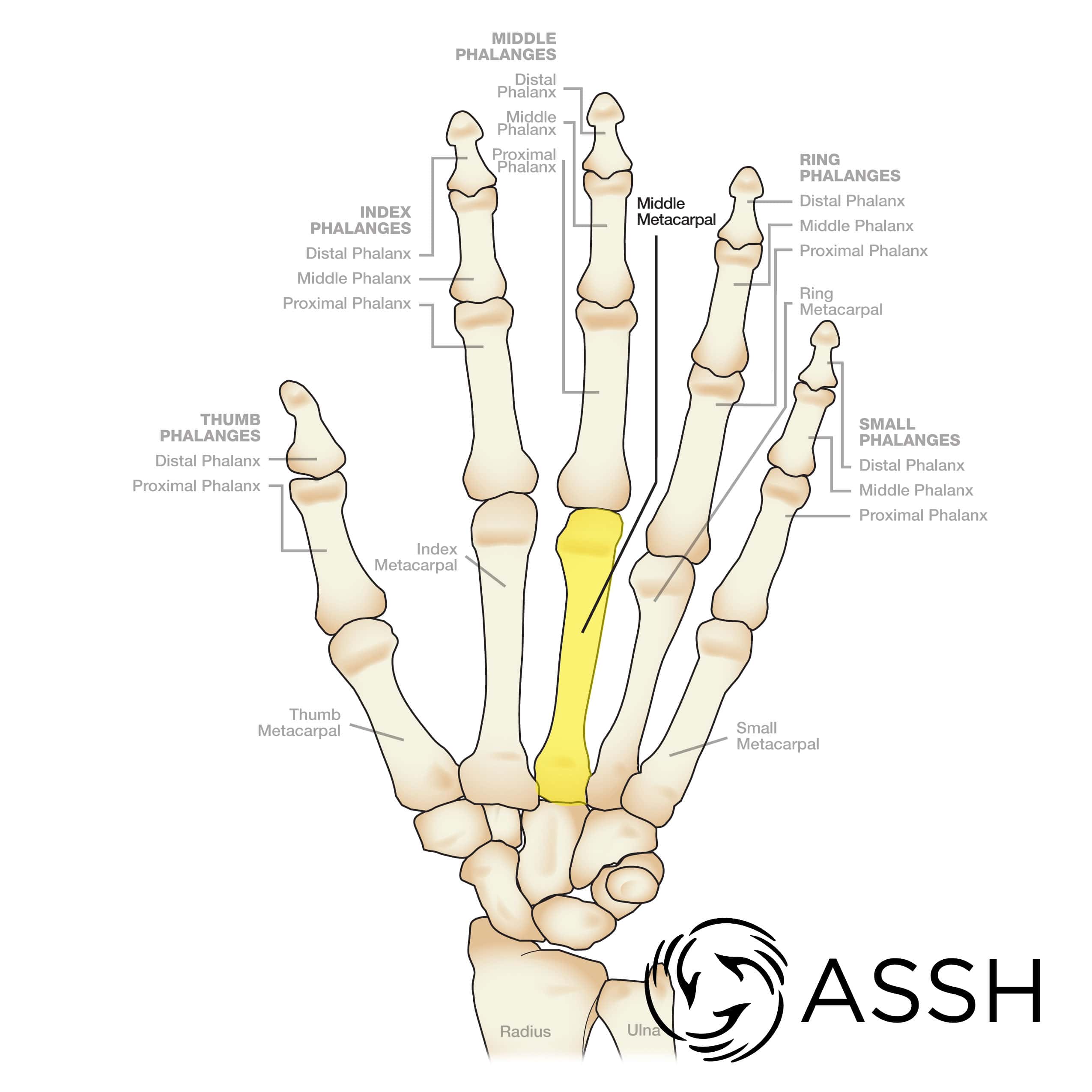
The metacarpals of the fingers make up the bone structure of most of the hand. They are all similar in shape and have joints in the wrist on one end, and the finger at the other end. The index and middle finger metacarpals have very little motion, while the metacarpals of the ring and little finger move much more.
Proximal phalanges
The proximal phalanx of the fingers is the proximal, or first bone, in the fingers when counting from the hand to the tip of the finger. There are three phalanges in each finger. The proximal phalanx is the largest of the three bones in each finger. The proximal phalanx has joints with the metacarpal and with the middle phalanx.
Middle phalanges
The middle phalanx of the finger is the middle or second of the three bones in each finger when counting from the hand to the tip of the finger. The middle phalanx has joints with the proximal phalanx and with the distal phalanx of the finger.
Distal phalanges
The distal phalanx of the finger is the distal or third of the three bones in each finger when counting from the hand to the tip of the finger. The distal phalanx has a joint just with the middle phalanx. On the tip of the phalanx is a bulbous tuft of bone that helps give the finger its rounded appearance. The distal phalanx is also important for supporting the fingernail.
Thumb metacarpal
The thumb metacarpal is similar in shape to the metacarpals of the fingers, but it is thicker. The thumb metacarpal has significantly more motion than the other metacarpals. It makes a joint with the trapezium that allows much of the thumb motion. This joint allows the thumb to move in a way that allows pinching. This is largely due to the unusual shape of the base of the metacarpal and the trapezium. The head of the metacarpal has a large joint surface next to the thumb proximal phalanx.
Thumb sesamoids
The thumb sesamoids are two small round bones at about the level of the thumb metacarpophalangeal joint These bones, as with all sesamoid bones, lie within tendons. The flexor pollicis brevis tendon and the adductor pollicis attach to the thumb sesamoids. Sesamoid bones help change the line of pull for their tendons which can help increase the force of tendon pull across the joint.
Thumb proximal phalanx
The thumb proximal phalanx is a short and stout bone between the metacarpal and distal phalanx. There is no middle phalanx in the thumb.
Thumb distal phalanx
The thumb distal phalanx is a short bone with a rounded tuft at the end that makes a joint with the proximal phalanx. The bulbous tuft at the end of the bone gives the thumb its rounded end. This bone supports the thumb nail.
Elbow Bones
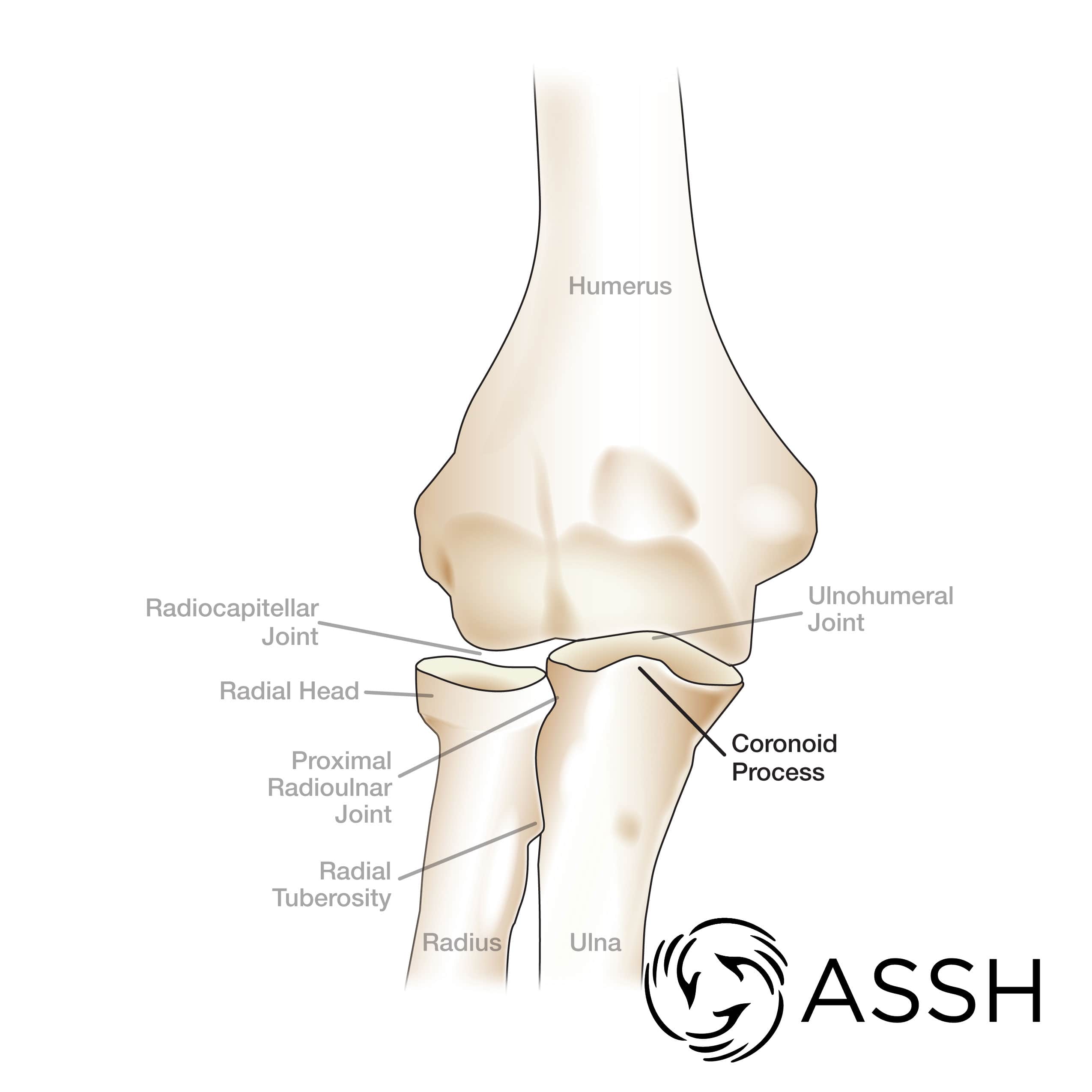 Coronoid process
The coronoid process is a small projection of bone off of the ulna bone that sits in the front of the elbow and on the inside of the elbow On and very near the coronoid process are attachment sites for muscles and ligaments of the elbow joint. The coronoid process is important for adding stability to the elbow.
Radial head
The radial head is a somewhat rounded cup that joins with the humerus and ulna to make up a portion of the elbow joint. The radial head has cartilage surfaces for both the humerus and the ulna to allow bending and extending of the elbow and twisting of the forearm. It also can add a significant amount of stability to the elbow joint.
Radial tuberosity
The radial tuberosity is a small, smooth projection on the surface of the radius bone near the elbow. It is the attachment site for the biceps tendon in the forearm. Because of the position of the tuberosity on the radius, the biceps tendon twists the hand palm up the forearm.
Lateral epicondyle
The lateral epicondyle is a bone projection off of the outside of the humerus bone. It is important primarily because of its soft tissue attachments to ligament and tendon.
Medial epicondyle
The medial epicondyle is a bone projection of the inside of the humerus bone. It is important primarily because of its soft tissue attachments to ligament and tendon. The ulnar collateral ligament and the common flexor tendon attach here. The ulnar nerve runs immediately behind the medial epicondyle.
Olecranon
The olecranon is a large projection of bone on the back of the elbow. It is a part of the ulna bone and makes the point of the elbow.